
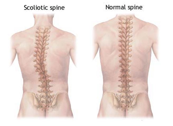
The human spine is made up of individual bones which are referred to as ‘vertebrae.’ The easiest way to consider the spine is to image these vertebrae stack on top of each other with a small cushion in between. This cushion is the intervertebral disc and provides shock absorption and flexibility. The spine is divided into three sections; cervical, thoracic and lumbar. Adolescent idiopathic scoliosis may present in any region of the spine and is seen as a curvature of the spine away from its normal alignment. While there are many forms of scoliosis, today we will specifically be exploring adolescent idiopathic scoliosis as it is perhaps the most common clinical presentation that we see in our clinic.Scoliosis is typically divded into three categories: congenital (from birth), idiopathic (unknown origin) and neuromuscular (deformity due to problems including Cerebral Palsy, Muscular Dystrophy, Spina Bifida and more). Adolescent idiopathic scoliosis is diagnosed when there is a persistent lateral curvature of the spine. This curvature is assessed via Chiropractic postural x-rays and may also involve rotation of the vertebrae.
Adolescent Idiopathic Scoliosis
Adolescent idiopathic scoliosis presents between the ages of 10yrs and 17yrs. The condition is more common in females and women who do present with the structural problem require ongoing monitoring as it’s common for it to progressively worsen over time. Usually the problem is painless, however associated muscle tightness and stiffness may occur. Typically it is discovered by a family member or an individual who notices that their clothes don’t fit like they used to.
Physical Examination
For all individuals who suspect they’re suffering from Adolescent idiopathic scoliosis it is important to seek appropriate help from a qualified and professional musculoskeletal specialist. Physical examination usually involves observing the spine for deviation and curvature as well as various orthopaedic tests such as the ‘forward bend test’ which demonstrates an obvious rib hump which can be measured. In combination with a thorough physical examination, postural spinal radiographs may be suggested by your healthcare practitioner. Spinal radiographs are important in such cases as it enables the practitioner to accurately measure the curve severity. This can be used as a baseline measure and re-evaluated if management is recommended.
Suggested Treatment Options
Suggested treatment options are largely influenced by the severity of the spinal curvature. Generally speaking it is recommended that:
- Curves 0° to 20° – manual therapy recommended or no management and assess for progression
- Curves 20° to 25° – manual therapy recommended or brace is advised if progression has been noted
- Curves 25° to 30° – manual therapy and referral to specialist
- Curves 30° and over – potential surgical intervention depending upon age and rate of progression
Recent Comments